
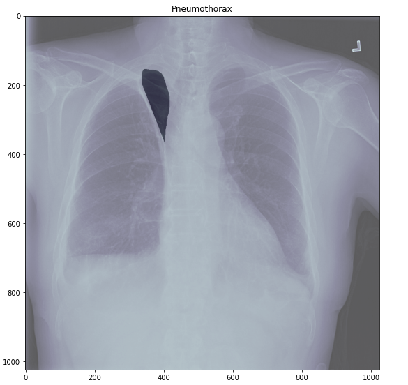
The primary objective of this task is identification and segmentation of chest radiographic images with pneumothorax.
Pneumothorax is usually diagnosed by a radiologist on a chest x-ray, and can sometimes be very difficult to confirm. An accurate AI algorithm to detect pneumothorax would be useful in a lot of clinical scenarios.
AI could be used to triage chest radiographs for priority interpretation, or to provide a more confident diagnosis for non-radiologists.
Definition: Pneumothorax can be caused by a blunt chest injury, damage from underlying lung disease.
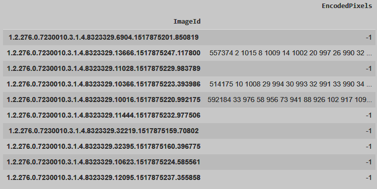
Data: Set of chest radiographic images in DICOM format(〜15.000 images) provided by Society for Imaging Informatics in Medicine
Dataframe
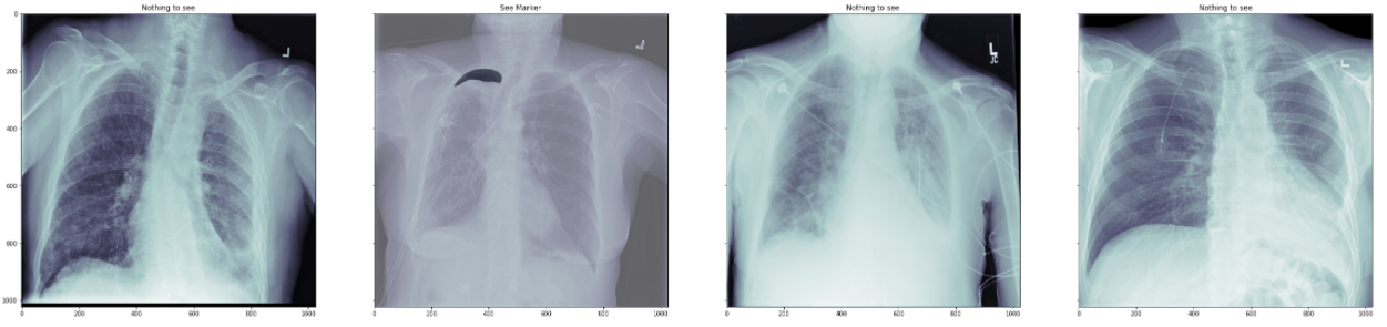
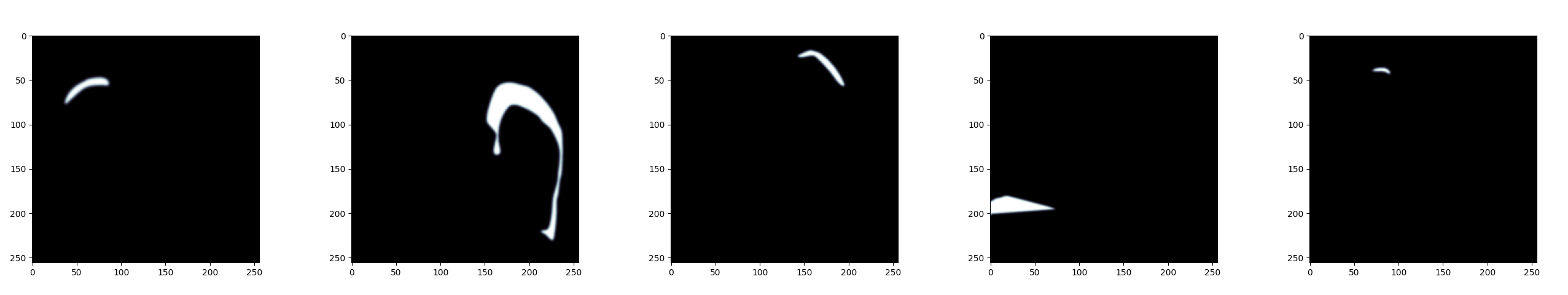
Data visualisation
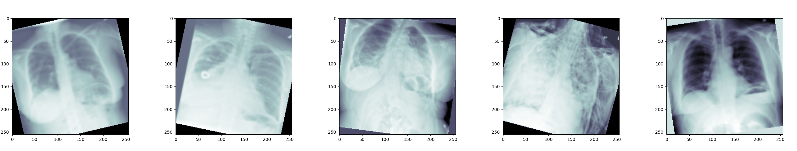
Data augmentation
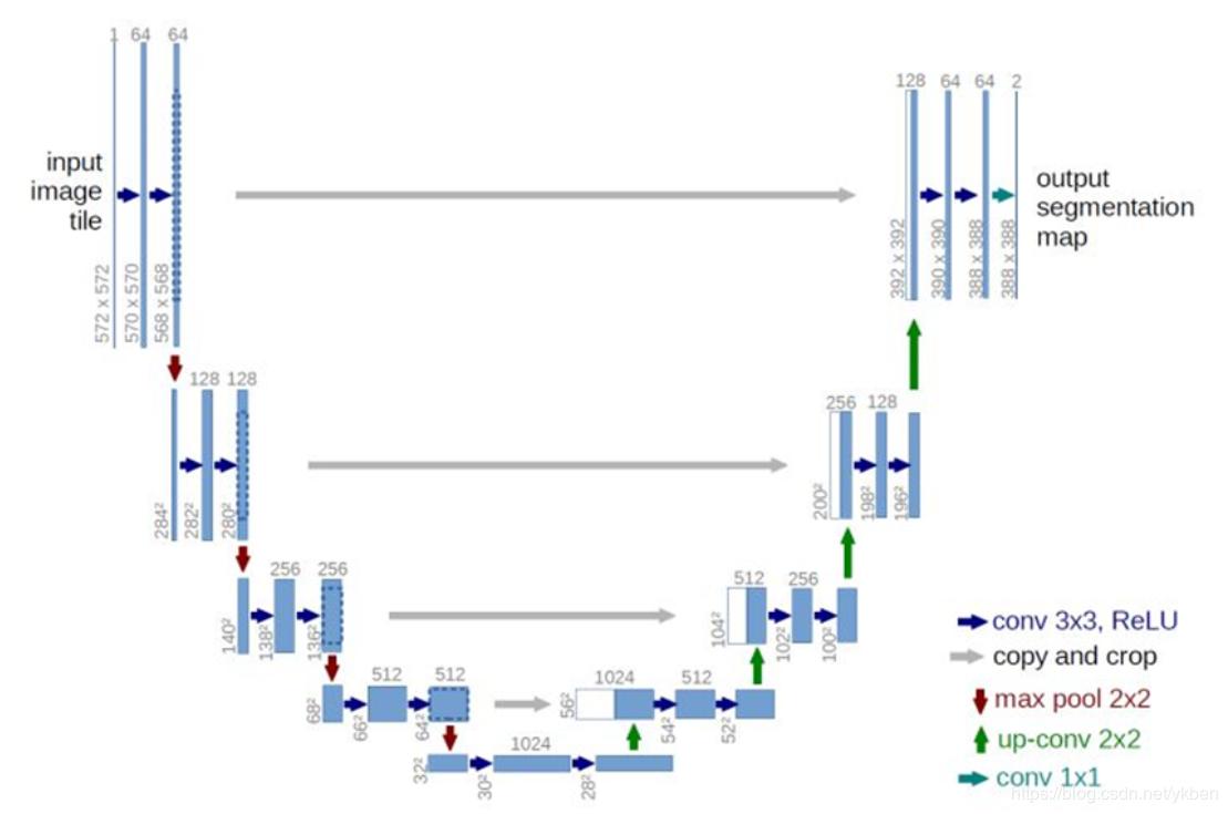
We used a U-net architecture with Adam as optimizer,
BCE with logistics loss and ReLU activation function.
The network was trained on 256 images for 130 epochs.
We used ROC and confusion matrix.
Calculated them from segmented masks of images with two classes ("Healthy" and
"Pneumothorax"). Their values are:
ROC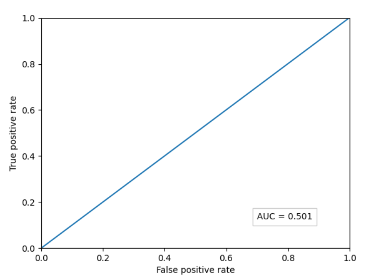
Confusion matrix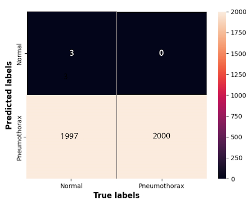
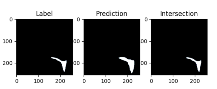
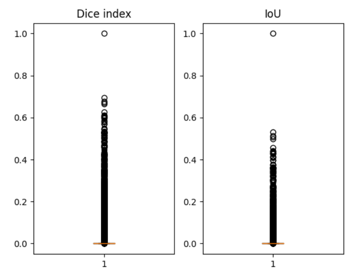
Comparing the true labels with the ground truth gave following
results:
Dice index: 0.043 +- 0.103
Intersection over union: 0.025 +- 0.065


P14 - Pneumothorax Segmentation for Summer School on Image Processing 2021