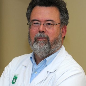
Erich Sorantin, MD, Ph.D.
Medical University of Grazpaediatric radiology, image processing, pattern recognition, 3D, radiation protection
Univ.-Prof. Dr.med.univ. Dr.h.c. Erich Sorantin was born in 1957 and, after finishing high school, he studied Medicine at the University of Vienna graduating in 1982. Afterwards he worked in several hospitals, fulfilled training as general practitioner and became fully qualified as Pediatrician and Radiologist. In 1988 he joined the team of the Department of Radiology, Medical University Graz, where he became a faculty member in 1994 and earned his professorship in 2002. Currently, Dr. Sorantin is the acting Head of the Division of Pediatric Radiology. Moreover, he coordinates a multi-institutional, multidisciplinary academic network in Central Europe, which focuses on biomedical imaging and technology transfer of advanced pediatric care as well as radiation protection. Additionally, Dr. Sorantin served as a consultant for Computer graphics for the medical vendor Siemens concentrating on virtual endoscopic techniques for approximately eight years. During this cooperation, two products were brought to the market: the Virtuoso3D and the Leonardo Workstation. Dr.Sorantin was decorated with several anwards and in 2018 he got a PhD honoris causa in Informatics from the Faculty of Science, Univ.of Szeged/HU. Dr. Sorantin has been married since 1984 and has three sons and two grandchildren.
Selected topics on biomedical image processing
Today’s imaging modalities like Multirow Detector Computed Tomography or Magnetic Resonance Tomography offer advanced geometrical and temporal resolution. Thus the generated data are already within the same range as the human genome. Therefore traditional reporting techniques like film or monitor reading are not longer appropriate. Moreover, the high computational power of recent workstations allows the implementation of more complex algorithms in order to assist the reporting radiologist, especially supporting quantitative tasks.
The aim of the lecture is to demonstrate selected applications of biomedical imaging and visualization regarding quantitative assessment of airway stenosis, computer aided diagnosis and virtual imaging with special emphasis on virtual endoscopic techniques as well as virtual operation planing.
Keywords: medical imaging, image processing, 3D