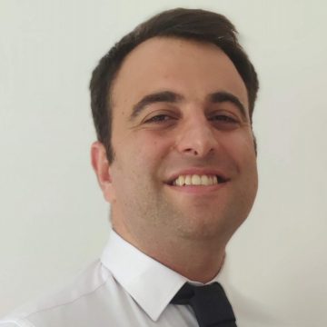
Ilkay Oksuz, Ph.D.
Istanbul Technical Universitymedical image segmentation, image registration, image quality assessment, cardiac MR, cardiac CT
Dr. Ilkay Oksuz is currently an Assistant Prof. in the Computer Engineering Department of Istanbul Technical University. His current research interests are in machine learning and deep learning, with a focus on medical image quality assessment, medical image reconstruction and segmentation. He has been working as a member of the King’s College London Biomedical Engineering Department under mentorship of Dr. Andrew King and Prof. Julia Schnabel since August 2017. He got his Ph.D. degree in Computer, Decision and Systems Science from IMT School for Advanced Studies Lucca, under supervision of Prof. Sotirios Tsaftaris. In 2017, he worked as a visiting researcher in IDCOM lab of The University of Edinburgh. In 2016, he was a postgraduate fellow at Diagnostic Radiology Department of Yale University under mentorship of Prof. Xenios Papademetris.
Medical image segmentation with topological constraints
Segmentation is the process of assigning a meaningful label to each pixel in an image and is one of the fundamental tasks in image analysis. Significant progress has been made on this problem in recent years by using deep convolutional neural networks (CNN), which are now the basis for most newly developed segmentation algorithms. Typically, a CNN is trained to perform image segmentation in a supervised manner using a large number of labelled training cases, i.e. paired examples of images and their corresponding segmentations. For each case in the training set, the network is trained to minimise some loss function, typically a pixelwise measure of dissimilarity (such as the cross-entropy) between the predicted and the ground-truth segmentations. However, errors in some regions of the image may be more significant than others, in terms of the segmented object’s interpretation, or for downstream calculation or modelling. Nonetheless, loss functions that only measure the degree of overlap between the predicted and the ground-truth segmentations are unable to capture the extent to which the large-scale structure of the predicted segmentation is correct, in terms of its shape or topology. In this talk, I will cover the neural network based segmentation methods that can incorporate topology and shape information for medical image segmentation. The examples will focus on computer assisted interventions and cardiac MRI myocardium segmentation.
Keywords: image segmentation, topology, shape, neural networks