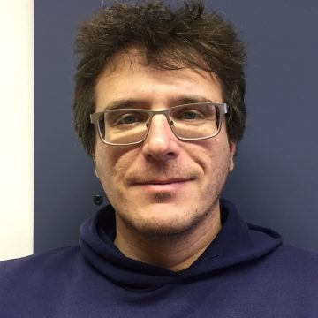
Matija Milanič, Ph.D.
University of Ljubljana, Faculty of Mathematics and Physics; Jozef Stefan Institutemedical imaging, spectroscopy, medical laser therapy, light transport modelling
Matija Milanic received the MSc/Diploma in Physics from University of Ljubljana (UL) in 2001. From 2001 to 2003 he was a teaching assistant for Technical physics at UL. From 2003 and 2008 he was a PhD student at Jozef Stefan Institute (JSI) working in the field of laser tissue interactions. In 2008 he received the PhD degree from UL for his thesis about pulsed photothermal radiometry. He continued his work in the field of laser medicine at JSI until 2013 including computational simulations of light-tissue interactions. From 2013 to 2015 he was a postdoc at NTNU Trondheim in the group of Lise L. Randeberg, where he was introduced to hyperspectral imaging (HSI) in medicine. The main research projects were application of HSI in rheumatology and detection of hypercholesterolemia. In 2015 he returned in Ljubljana, where is an assistant professor in Medical physics at University of Ljubljana and a researcher at IJS. He is a member of APS, group on Medical physics, MC member at COST actions focusing on medical therapy and diagnostics, and since 2020 an expert for European Commission’s expert panels on medical devices and in vitro diagnostic medical devices.
His current research interests are light–tissue interactions and translation of this knowledge to clinical and industrial environment. He is involved in exploration of spectral properties of light for diagnostic purposes (e.g., spectral imaging of joint inflammation, peritonitis, skin lesions) and understanding of tissue physiology and morphology (e.g., tissue oxygenation, scattering, chromophores distribution). In addition to the medical applications he is also involved in applications of spectral imaging in food science and cultural heritage.
Processing of spectral images
Spectral imaging is imaging where images of an object or a scene are recorded at more than one spectral band. A common example of spectral imaging is RGB imaging where images are acquired at three different spectral bands: red, green and blue. However, spectral images can have a lot of different spectral bands – in case of hyperspectral imaging few hundreds or thousands bands. Because of the combination of spatial and spectral information, spectral images are typically processed using a different algorithms as monochromatic images.
In this lecture I will focus on overview of the spectral imaging technique and technology, typical spectral image processing pipeline including pre-processing (data normalisation, noise reduction) and processing (feature extraction, classification), all illustrated by examples from medicine, cultural heritage and food quality inspection. At the end, hot-topics in spectral imaging will be presented.
Keywords: multispectral, hyperspectral, medical imaging, cultural heritage